
Figure 5.
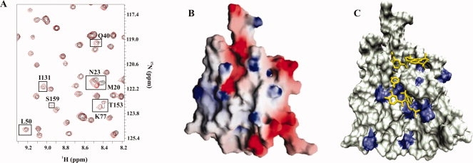
Suramin binding to PA1324. (A) An expanded region of an overlay of the 2D 1H-15N HSQC spectrum of free PA1324 (black) and PA1324 in the presence of suramin (red). (B) A GRASP40 electrostatic surface of PA1324 demonstrating the positive electrostatic potential of the suramin binding site. Blue and red indicate positively charged and negatively charged surfaces, respectively. (C) A docked model of suramin in the PA1324 binding site identified by FAST-NMR. Residues that incurred a CSP upon binding suramin are colored blue. The distal CSPs could be long-range effects of the binding interaction or potential suramin conformational exchange.