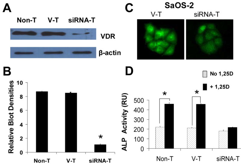
Fig. 2.

Stable VDR silencing in SaOS-2 cells. (A) VDR protein expression levels in native non-transfected (Non-T), control (vector transfected, V-T), and siRNA VDR transfected (siRNA-T) Saos-2 cells. VDR levels are quantitatively shown in (B) as a measure of blot densities. (C) Immunofluorescence detection of VDR in control V-T (left), and VDR silenced (siRNA-T, right). Cells treated with a goat antibody against human VDR were visualized with a secondary anti-goat Cy3-conjugate antibody. VDR was profusely localized in the cell cytoplasm and nucleus of control SaOS-2 cells. Note significantly decreased fluorescence intensity levels corresponding with lower VDR protein levels in VDR silenced versus control V-T cells. Digital images were obtained with identical exposure settings for comparison. (D) 1,25D induction of alkaline phosphatase (ALP) activity requires a VDR. Endogenous ALP enzyme activity was measured in the absence (open bars) and presence (filled bars) of 10 nM 1,25D added to the culture medium for 3 days in native Non-T, control V-T, and siRNA VDR transfected SaOS-2 cells. *, p<0.001, n=3.
