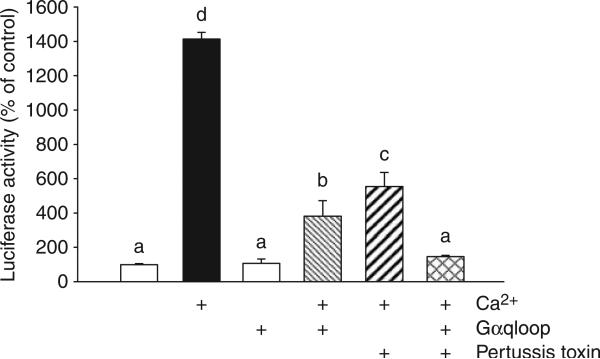
Figure 1. CASR is coupled to activation of Gαq and Gαi.
HEK-293 stably CASR-expressing cells were cotransfected with the constructs directing the expression of Gαqloop (corresponding to residues 305−359 of mouse Gαq; 1.5 μg), the SRE-luciferase reporter gene (0.5 μg) and p-cytomegalovirus-β-galactosidase (pCMV-β-gal; 0.015 μg) per well of six-well plate. The transfection is described in ‘Materials and Methods’. After 2 days of transfection, quiescent cells were pretreated with vehicle or 100 ng/ml pertussis toxin for 5 h, then stimulated by 5 mm calcium for the last 8 h. Data are shown as relative luciferase activity reported as the percent induction, compared with the activity under nonstimulated conditions and normalized for β-galactosidase. Values represent the mean±s.e.m. of at least three experiments. Values sharing the same superscript are not significantly different at P≤0.05.

