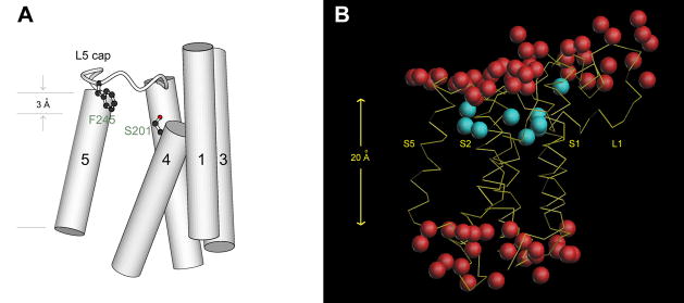
Fig. 5.
Features of the high-resolution crystal structure of E. coli GlpG. (A) The catalytic serine is located 3 Å below the membrane surface, and points up. (B) The α-carbon trace of the protein backbone is shown in yellow. Waters bound inside the protein are shown in cyan. The externally bound waters, observed in the 1.9 Å resolution crystal structure (PDB entry 3b45) and shown in red, are found in two layers, which roughly correspond to the boundaries of the membrane.

