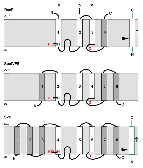
Fig. 7.
The membrane topology models for RseP, SpoIVFB and S2P (substrates shown in blue). The arrow head points at the cleavage site; the arrow indicates the direction of the polypeptide; the metal binding motifs are shown in red. The conserved three TM domains (a–c) are represented by white boxes. An 8-TM model of S2P is shown here (see text). Note that the figure is not in scale; the various “loops” that connect the TM domains may fold into α-helices (or even continuous with the TM helix) or β-strands in the real structure.

