
Figure 3.
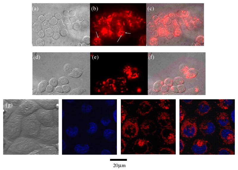
Fluorescence images of KB cells incubated with dual-functionalized DNA-NTs for one hour. (a-c) before and (d-f) after extensive washing. (a) & (d): brightfield images; (b) & (e): fluorescence images; (c) & (f): the superimposed images of (a) & (b) and (d) & (e), respectively. White arrows in (b) indicate some recognizable DNA-NTs. (g) Confocal fluorescence images of washed cells. From left to right: brightfield image, Hoechst stain (indicating the nuclei) image, Cy3 (indicating DNA-NTs) image, superimposed nuclei and DNA-NT image. Hoechst 33342: a blue fluorescence dye that specifically stains nuclei.