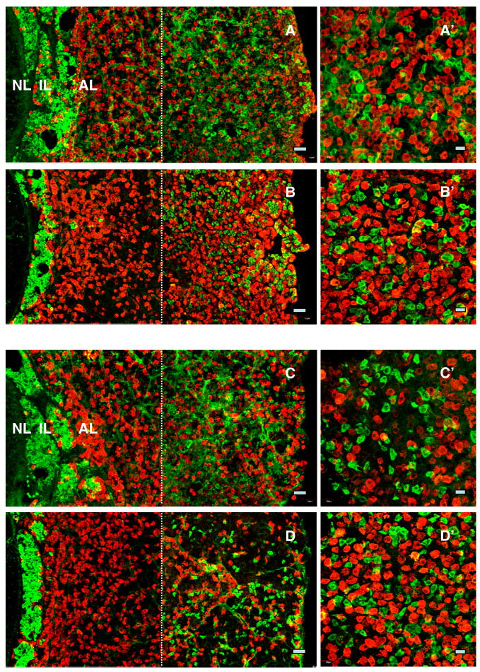
Figure 4.
Confocal projections (20X; 1 μm optical sections) of dual immunofluorescence images of kisspeptin cells (green fluorescence, Alexa 488) in relation to LH (panels A and B) and FSH (red fluorescence, Cy3, panels C and D) immunopositive cells in pituitary from an intact (panels A and C) and a castrated (panels B and D) monkey. Kisspeptin cells were distributed in the intermediate lobe (IL) and towards the periphery of the anterior lobe (AL), whereas gonadotropin immunopositive cells were found throughout the AL. Absence of colocalisation of kisspeptin with either LH (A′ and B′) or FSH (C′ and D′) secreting cells is shown in higher magnification confocal projections (40X; 1 μm optical sections). In cases where orange fluorescence was observed, examination of individual optical 1 μm sections revealed that the merged optical signal was due to the adjacent location of kisspeptin cells and gonadotrophs. Note the left hand photomicrographs were again generated by creating a montage of two images separated by the thin broken white vertical line: the left hand image captured the NL, IL and AL immediately adjacent to IL, while the right hand image shows peripheral AL. Scale bars, 50 μm (A–D) and 20 μm (A′–D′).

