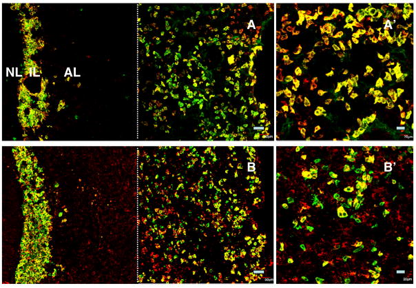
Figure 6.
Confocal projections (20X; 1 μm optical sections) of dual immunofluorescence images of kisspeptin (green fluorescence, Alexa 488) and α-MSH immunopositive cells (red fluorescence, Cy3) in the intermediate lobe (IL) and in the anterior lobe (AL) of the pituitary from an intact (A) and a castrated (B) monkey. The majority of cells in the IL (melanotrophs) expressed both peptides (A and B). Colocalisation of kisspeptin and α-MSH was also observed in peripheral areas of AL (A and B). This double label staining of presumptive corticotrophs is also shown in higher magnification (40X; 1 μm optical sections) in panels A′ and B′. Note the left hand photomicrographs were again generated by creating a montage of two images as described in previous figures. Scale bars, 50 μm (A and B) and 20 μm (A′, B′).

