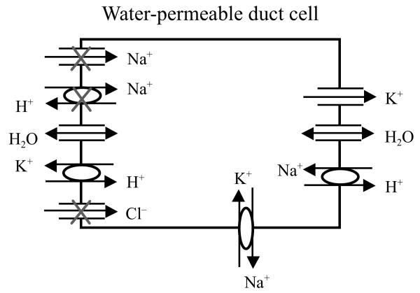
Fig. 1.
Schematic diagram of the hypothesized mechanism for fluid secretion following AdhAQP1-mediated gene transfer to duct cells in an irradiated salivary gland. The surviving duct cell is presented in a simplified form, based on our understanding ∼1991. The duct lumen is to the left and the interstitium is to the right. The three apical ion pathways with Xs will be inoperable in the absence of an isotonic primary secretion, which normally is made by acinar cells. We have hypothesized that duct cells could generate a KHCO3 gradient (lumen > interstitium) enabling fluid to flow into the duct lumen following expression of the transgene hAQP1. This figure is modified from Vitolo and Baum (2002), and is based on the experiments presented in Delporte et al. (1997)

