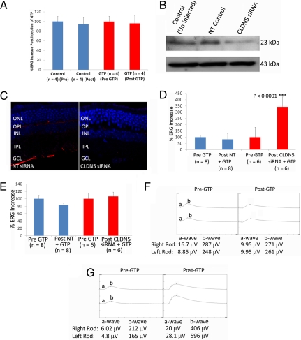
Fig. 3.
Enhanced delivery of GTP to retinas of IMPDH1−/− mice. (A) After analysis of ERG readout in IMPDH1−/− mice, animals were injected with a 3-μL solution containing 0.6 mg of GTP. Mice were again analyzed by ERG, and the percentage change in electrical readout from the retina was plotted. There was no significant difference in ERG readouts in IMPDH1−/− mice after intraocular injection of GTP (n = 4 mice per group). (B) Levels of expression of claudin-5 in the retinas of IMPDH1−/− mice were assessed by Western blotting, with levels decreased 48 h after injection of claudin-5 siRNA. (C) From immunohistochemical analysis of claudin-5 levels (red staining) in retinal cryosections, it was clear that levels were decreased in all retinal layers. Sections were counterstained with DAPI (blue staining). (40× objective.) (D) IMPDH1−/− mice were subjected to ERG analysis of rod function, and 48 h after injection of either an NT or claudin-5 siRNA, mice were administered an injection of GTP, and ERG readout was assessed again. Electrical readout of rod photoreceptors was expressed as percentage changes, and a significant increase (***, P < 0.0001) in rod-isolated ERG was observed in mice receiving claudin-5 siRNA and 3.3 mg of GTP compared with mice receiving NT siRNA and 3.3 mg of GTP. (E) No change in retinal cone function was observed after injection of GTP in either experimental group. (F) Rod-isolated ERG tracings in IMPDH1−/− mice before and after injection of GTP with NT siRNA showed no distinct changes in waveform; however, upon analysis of ERG tracings before and after GTP injection with claudin-5 siRNA, well-formed a and b waves were observed in the retinas of mice.

