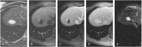
FIGURE 1.
Typical hemangioma in a 41-year-old woman. T2-weighted image (TR/TE, 5000:100 milliseconds) with fat suppression shows a hyperintense lesion in the right lobe (A). Transverse dynamic fat-suppressed T1-weighted MR images (TR/TE, 5.1:1.2 milliseconds) of this lesion show peripheral nodular enhancement in the hepatic arterial phase (B), centripetal enhancement in the portal venous phase (C), progressing into complete uniform filling in the delayed phase (D). Diffusion-weighted image (b = 500; TR/TE, 6500:110 milliseconds) shows a bright lesion with an ADC value of 2.14 × 10⁻³ mm2/s (E).

