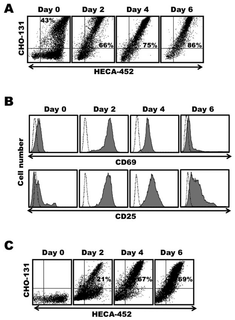
Figure 2. Memory CLA+ CD4+ T cells are stained in essentially a uniform manner by CHO-131 following their activation.
Freshly isolated, peripheral blood lymphocytes sorted for CLA+ CD4+ T cells or CHO-131− CLA+ CD4+ T cells were stimulated with anti-CD3 mAb plus IL-2, as described in Figure 1. Day 0, freshly isolated cells; Day 2, activation phase; Days 4 and 6, expansion phase. (A) Activated CLA+ CD4+ T cells were harvested at the indicated time points and dual stained with the mAbs CHO-131 and HECA-452. (B) In addition, the activated CLA+ CD4+ T cells were stained for CD69 expression (top panels) or CD25 expression (bottom panels). (C) The expression kinetics of C2-O-sLeX by activated CHO-131− CLA+ CD4+ T cells was analyzed by dual staining with the mAbs CHO-131 and HECA-452. All analyses were performed by flow cytometry. For all plots, the indicated mAb reactivities on the x and y axes represent Log 10 fluorescence. Non-specific antibody labeling was determined using the appropriate isotype negative control antibodies, as indicated and data not shown. Data are representative of at least three independent experiments using T cells isolated from separate donors.

