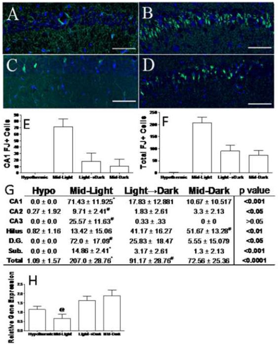
Figure 3. Time-of-day determines that cell death responses to ischemia.
Representative sections of Fluoro-jade C (degenerating neurons; green) and DAPI (nuclear counterstain; blue) stained tissue from the CA1 region of mice that underwent hypothermic CA/CPR (a) or normothermic CA/CPR during the Mid-Light (b), Light-Dark transition (c), or Mid-Dark period (d). Quantification of Fluoro Jade positive neurons in the CA1 field (e) and across the whole hippocampus (f). Table of mean (±SEM) for Fluoro-jade positive neurons in selected regions (g). Hippocampal bcl-2 gene expression relative to 18s rRNA gene expression at 24 h post-CA/CPR (h). Scale bar =100μm. CA -Cornu Ammonis, D.G. dentate gyrus, Sub. subiculum. * indicates significantly different from all other groups at p<0.05; # indicates significantly different from hypothermic mice at p<0.05. n=7–11/group for histology and 4–10/group for PCR.

