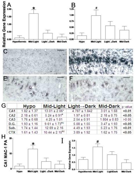
Figure 4. Time-of-day determines inflammatory responses to cardiac arrest.
Proinflammatory cytokine gene expression was potentiated by CA/CPR during the Mid-Light. (a) Il-1β and (b) Tnfα mRNA gene expression relative to 18s rRNA. Microglial activation was also potentiated in mice that underwent ischemia during the middle of the light period. Representative sections of MAC-1 stained sections through the CA1 field of the hippocampus in mice that underwent hypothermic CA/CPR (c) or normothermic CA/CPR in the Mid-Light (d), Light→ Dark Transition (e) or in the Mid-Dark (f). Table of proportional areas of microglial activation across the hippocampus and associated regions (g). (h) MAC-1 proportional area in the CA1 field. (i) Immunohistochemical evidence of increased microglial activation was confirmed with MAC-1 rat-PCR 24 h post-CA/CPR. Data are presented as the mean ±SEM. * significantly different from all other groups (p<0.05); # significantly different from hypothermic mice (p<0.05); c significantly different from mid-dark mice (P<0.05); d significantly different from Light→Dark mice (p<0.05). Scale bar =100μm. CA-Cornu Ammonis, D.G. dentate gyrus, Sub. subiculum, CTX, posterior parietal cortex n=7–11/group for histology and 4– 10/group for PCR.

