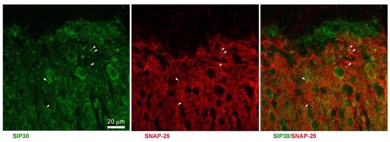
Fig. 7.
Double immunofluorescence staining shows that SIP30 (green) co-localized with SNAP-25 (red) in the dorsal horn of the spinal cord. Left panel: SIP30 immunoreactivity was present in both soma and terminals of spinal dorsal horn neurons. Middle panel: SNAP-25 immunoreactivity was present in presynaptic terminals of spinal dorsal horn neurons. Right panel: SIP30 (green) co-localized with SNAP-25 (red) in terminals of spinal dorsal horn neurons. Arrowheads indicate double-labeled terminals.

