Figure 4.
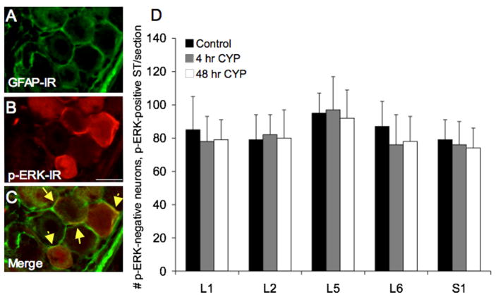
Pericellular pERK1/2-IR in lumbosacral DRG is present in control and CYP-treated rats and does exhibit regulation with cystitis. Pericellular pERK1/2-IR appeared to be present in satellite cells surrounding L6-S1 DRG cells. A–C: L6 DRG section exhibiting pericellular pERK1/2-IR (red; B) was also stained for glial fibrillary acidic protein (GFAP; green; A). C: Merged image of A and B displaying some areas of overlap (yellow arrows) between pericellular pERK1/2-IR and GFAP consistent with pERK1/2 expression in satellite cells. Calibration bar represents 20 μm in A–C. D: Summary histogram of numbers of pERK1/2 cytoplasmic negative DRG neurons-pERK1/2 pericellular positive satellite (ST) cell staining in DRG. Pericellular pERK1/2-IR in L1, L2, L5-S1 DRG was not regulated by CYP-induced cystitis (4 hr or 48 hr). Data are a summary of n = 6–8 animals for each group.

