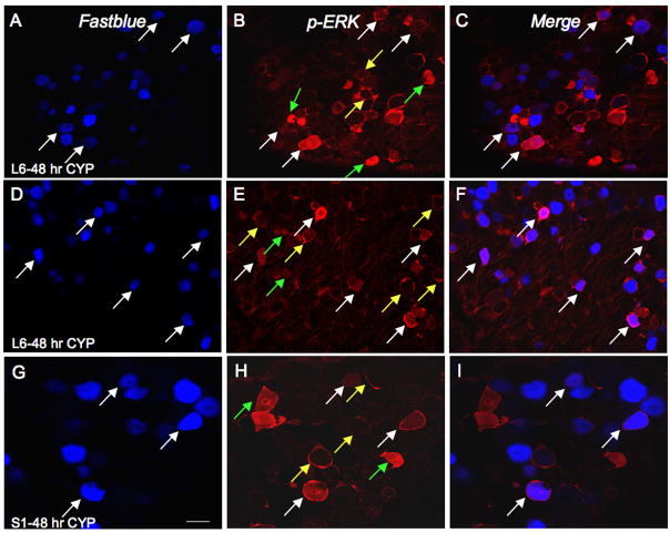
Figure 5.
CYP-induced cystitis increases pERK1/2-IR in bladder afferent (Fastblue, FB-labeled) cells in the lumbosacral DRG. A–C: L6 DRG section from rat treated with CYP (48 hr) showing bladder afferent cells (FB-labeled; A; white arrows) demonstrating pERK1/2-IR (B) in same section and merged image (C) demonstrating bladder afferent cells with pERK1/2-IR (pinkish-purple/magenta; white arrows). D–F: Another example of an L6 DRG section from rat with 48 hour induced cystitis showing bladder afferent cells (FB-labeled; A; white arrows) demonstrating pERK1/2-IR (B) in same section and merged image (C) demonstrating bladder afferent cells with pERK1/2-IR (pinkish-purple/magenta; white arrows). G–I: Higher power image of S1 DRG section from rat treated with CYP (48 hr) showing bladder afferent cells (FB-labeled; A; white arrows) demonstrating pERK1/2-IR (B) in same section and merged image (C) demonstrating bladder afferent cells with pERK1/2-IR (white arrows). Not all cells expressing pERK1/2-IR are bladder afferent cells (B, E, H; green arrows) and 25–29% of bladder afferent cells expressed pERK1/2-IR after 4 or 48 hr CYP-induced cystitis. Pericellular pERK1/2-IR cells are also indicated (yellow arrows); very few of these cells were bladder afferent cells. Calibration bar represents 40 μm in A–C, D–F; 20 μm in G–I.

