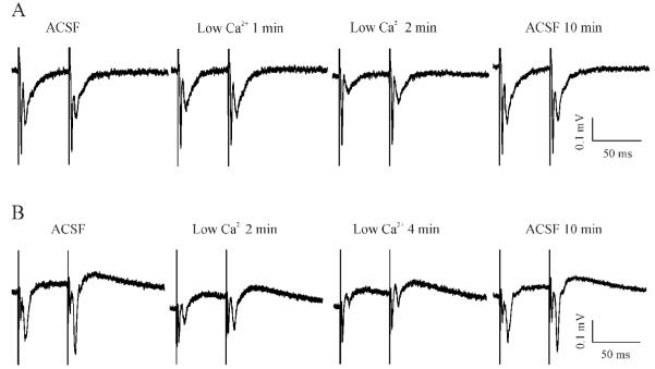
Figure 5.
Reducing extracellular Ca2+ concentrations changed the STP form in control slices but not MeHg-exposed slices. (A) A slice prepared from a rat with injections of 0.9% NaCl for 30 days showed a typical PPD response. Data are shown at selected time points when the slice was incubated in the standard 2 mM Ca2+-containing ACSF (ACSF), in a low Ca2+-containing solution for 1 or 2 min (Low Ca2+ 1 or 2 min), and in standard ACSF again for 10 min (ACSF 10 min). (B) Under the similar recording conditions as described for A, the slice prepared from a rat with injections of 0.75 mg/kg/day MeHg for 30 days demonstrated a typical PPF when incubated in the standard ACSF. Similarly, data are shown at selected time points when the slice was incubated in the standard ACSF (ACSF), in a low Ca2+-containing solution for 2 or 4 min (Low Ca2+ 2 or 4 min), and in standard ACSF again for 10 min (ACSF 10 min). Each trace is representative of 6 - 7 individual experiments.

