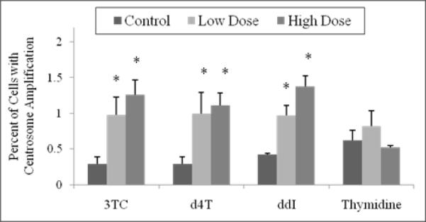
Figure 2. Percentage of CHO cells exhibiting centrosome amplification.
Cells were incubated with 12.2 μM and 122 μM 3TC, 8.9 μM and 89 μM d4T, 10.2 μM and 102 μM ddI, and 9.9 μM and 99 μM thymidine for 24 hr. Values are based on observation of 1000 cells. The stars indicate groups that are significantly different (p < 0.05) compared to unexposed control cells.

