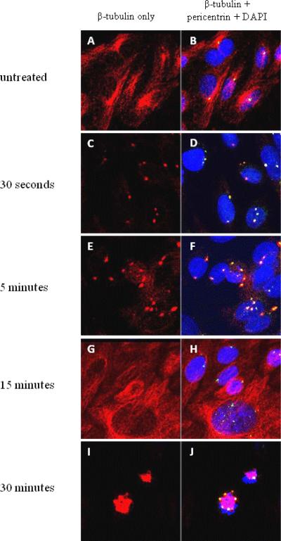
Figure 4. Microtubule nucleation of CHO cell centrosomes.
CHO cells incubated with an anti-pericentrin antibody (green), an anti-β-tubulin antibody (red), and DAPI for DNA (blue). (Colocalization of pericentrin and β-tubulin appears yellow and colocalization of pericentrin, β-tubulin, and DAPI appears white.)
(A, B): Untreated CHO cells, without nocodazole incubation.
(C, D): 30 seconds after nocodazole removal. Centrosomes begin to recover microtubule aster formation.
(E, F): 5 minutes after nocodazole removal. Microtubule asters grow larger.
(G, H): 15 minutes after nocodazole removal. Normal microtubule distribution.
(I,J): 30 minutes after nocodazole removal. Cell division with multi-polar spindles.

