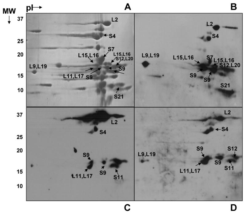
Fig. 1. Two-dimensional gel analysis of phosphorylated E. coli ribosomal proteins.

(A) 15 A260 units of E. coli ribosomes were separated on NEPHGE gels using pI 5-7 and 3-10 ampholytes. The gel was stained with Coomassie Blue. (B, C, D) Immunoblotting analysis of E. coli ribosomal proteins using phosphotyrosine, phosphoserine, and phosphothreonine antibodies, respectively. The regions labeled correspond to phosphorylated proteins identified by LC-MS/MS analysis from the tryptic digests of the gel pieces. Not all of the proteins that are found to be phosphorylated are marked in the figures.
