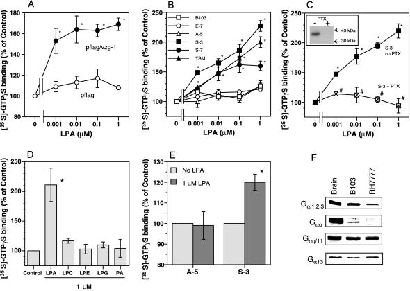
Figure 2.
VZG-1 directly couples to Gi and other G proteins in plasma membranes after LPA stimulation. (A) Effect of LPA on [35S]-GTPγS binding to membranes from RH7777 cells transiently transfected with pflag or pflag/vzg-1. Basal [35S]-GTPγS binding (no LPA) was 64.8 ± 5.3 (pflag) and 100.7 ± 6.8 (pflag/vzg-1) cpm/μg protein. Data are expressed as a percentage of control (no LPA) and represent the mean ± SEM (n = 3–4). ∗, P < 0.05 vs. no LPA (using Student’s t test). (B) Effect of LPA on [35S]-GTPγS binding to membranes of control and experimental B103 stable cell lines and cortical neuroblast cell line TSM. Basal [35S]-GTPγS binding was 45.7 ± 2.5 (B103), 61.9 ± 2.1 (E-7), 57.2 ± 4.2 (A-5), 72.6 ± 3.7 (S-3), 73.9 ± 5.5 (S-7), and 83.3 ± 5.8 (TSM) cpm/μg protein. Data are expressed as described in A (n ≥ 3). ∗, P < 0.05 (using Student’s t test) vs. no LPA. (C) Effect of PTX treatment on LPA-induced [35S]-GTPγS binding in clone S-3. Cells were treated without or with PTX. Basal [35S]-GTPγS binding was 72.6 ± 3.4 (no PTX) and 71.1 ± 1.3 (PTX) cpm/μg protein. (Inset) Autoradiogram of PTX-catalyzed [32P]-ADP ribosylation in membranes of S-3 cells pretreated without (Left) or with PTX (Right) demonstrates PTX functionality. Data are expressed as described in A (n = 3). ∗, P < 0.05 (using Student’s ttest) vs. no LPA. #, P < 0.05 vs. no PTX. (D) Effect of structurally related phospholipids on [35S]-GTPγS binding in clone S-3. Basal [35S]-GTPγS binding was 91.9 ± 20.2 cpm/μg protein. Data are expressed as described in A (n = 3). ∗, P < 0.05 (using Student’s t test) vs. no LPA. (E) VZG-1 couples to Gi after LPA stimulation. A-5 or S-3 cell membranes were labeled with [35S]-GTPγS, and Gαi protein was immunoprecipitated as described in Experimental Procedures. Basal binding in the immunoprecipitates was 15.2 ± 4.7 (A-5) or 20.0 ± 6.9 cpm/μg protein (S-3). Data are expressed as described in A (n = 3). ∗, P < 0.05 (using Student’s t test) vs. no LPA. (F) Western blot demonstrates expression of G proteins thought to mediate LPA signaling in mouse brain, B103, and RH7777 cell lines. Isolated membranes (50 μg) were subjected to Western blotting, as described (13). See Table 1 and Experimental Procedures for abbreviations.

