Figure 3.
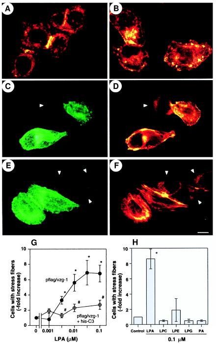
VZG-1 mediates stress fiber formation by LPA in RH7777 cells through Rho activation. Immunofluorescent microscopy of RH7777 cells transfected with pflag (A and B) or pflag/vzg-1 (C to E) and then exposed to LPA at 0 nM (A, C, and D) or 100 nM (B, E, and F) for 15 min. Cells were fixed and double-labeled for flag-epitope (using M2 antibody visualized by indirect immunofluorescence using fluorescein isothiocyanate; C and E) and actin (visualized with TRITC-phalloidin; A, B, D, and F). Note that stress fibers are observed only with VZG-1 expression and LPA exposure (E and F). Arrowheads, nontransfected cells. (Bar = 10 μm.) (G) Effect of his-C3 exoenzyme on LPA-induced stress fiber formation in VZG-1-expressing cells. RH7777 cells transfected with pflag/vzg-1 were treated without or with his-C3 exoenzyme, exposed to varying concentrations of LPA, and double-labeled for flag epitope and actin. The change in the number of double-labeled cells with stress fibers after LPA exposure compared with control (no LPA) was expressed as “fold increase.” Data are the mean ± SEM (n = 4). ∗, P < 0.05 (using Student’s t test) vs. no LPA. #, P < 0.05 vs. no C3 exoenzyme. (H) Effect of structurally related phospholipids on VZG-1-mediated stress fiber formation in pflag/vzg-1-transfected RH7777 cells. Data are the mean ± SEM (n = 3). ∗, P < 0.05 (using Student’s t test) vs. no LPA. See Experimental Procedures for abbreviations.

