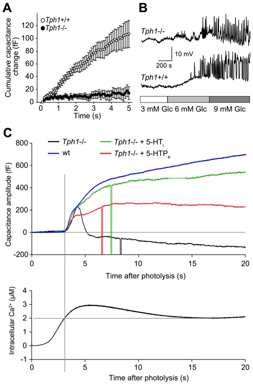
Figure 3. Impaired exocytosis of Tph1−/− β-cells and rescue with 5-HT.
(A) A train of 50 depolarizing pulses from −80 to +10 mV for 40 ms at 10 Hz induced changes in the membrane capacitance of wt (n = 24) and Tph1−/− (n = 19) β-cells. Data are shown as means ± SEM. (B) Representative current-clamp recordings of the electrical activity of wt and Tph1−/− β-cells in pancreas tissue slices. The slices were perfused with solutions containing different glucose concentrations as indicated in the bar below the traces. (C) Representative membrane capacitance response of isolated wt and Tph1−/− β-cells (top panel) stimulated by ramp [Ca2+]i change induced by slow photo-release of caged Ca2+ (bottom panel). The impaired component exocytosis has been partially rescued by extracellular 5-HTP (500 µM, 24 h) and completely restored by pipette intracellular dialysis with 5-HT.

