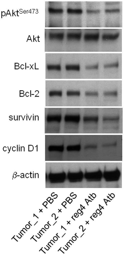
Figure 7. Expression of phosphorylated AKT, Bcl-xL, Bcl-2, surviving and cyclin D1 in pancreatic tumors treated with a specific anti-REG4 antibody.
Tumor tissue samples were lysed in a homogenization buffer containing a cocktail of antiproteases. Cell debris and insoluble materials were eliminated by centrifugation and soluble fractions were loaded in Laemmli buffer onto a 12.5% SDS-polyacrylamide gel. The proteins were transferred to nitrocellulose membrane and membrane incubated with the anti phosphorylated AKT (phospho-Ser473), anti-pan AKT, anti-Bcl-2, anti-Bcl-xL, anti-survivin or anti-cyclin D1 antibodies. After development, the membrane was stripped and reprobed for β-actin.

