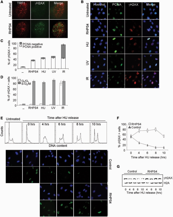
Figure 1.
Replication-dependent induction of damage by RHPS4. (A) BJ-EHLT fibroblasts treated with RHPS4 for 16 h were fixed and processed for IF by using antibodies against γH2AX and TRF1. Representative deconvolution images are reported. (B) BJ-HELT fibroblasts were exposed to the following treatment: 0.5 μM RHPS4 for 16 h, 2 mM HU for 3 h, UV 10 J/m2, IR 5 Gy. Representative images of IF against γH2AX and PCNA were acquired with a Leica Deconvolution microscope (magnification: ×40). (C) Percentage of γH2AX+/PCNA- or γH2AX+/PCNA+ nuclei in the indicated samples. The mean of three independent experiments with comparable results is shown. (D) HeLa cells untreated or exposed to the indicated treatment were sorted by FACS according to the DNA content. The fractions corresponding to cells in G0/G1 and S–G2/M cell cycle phases were cytocentrifuged on cover slips and stained for IF against γH2AX. Histogram represents the percentage of γH2AX-positive cells under different stimulations. The mean of three independent experiments with comparable results is shown. (E) BJ-HELT fibroblasts were treated with a low dose of HU (0.5 mM for 16 h) to block cells at the G1–S boundary. Then, the medium was replaced to release cells and in the treated samples 0.5 μM RHPS4 was added. Cells were fixed at the indicated times for cell cycle analysis (upper panel) and IF against γH2AX. Representative images of IF are reported in the lower panel (magnification: ×63). Percentage of γH2AX-positive nuclei (F) and western blotting analysis of γH2AX (G) in control and RHPS4-treated samples at different times after HU release.

