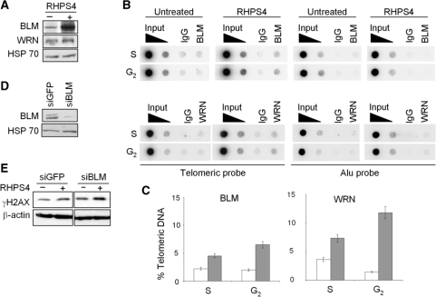
Figure 4.
BLM and WRN helicases are increased and recruited to the telomeres upon RHPS4 treatment. (A) Western blot analysis of BLM and WRN helicases in BJ-HELT cells untreated or treated with RHPS4 for 96 h. (B) ChIP experiments on BJ-HELT fibroblasts untreated and treated with RHPS4. Protein extracts from cells during S and G2 phases of cell cycle were subjected to ChIP experiments using antibodies against BLM and WRN. IgG antibody was used as negative control. The total DNA (input) represents 10 and 1% of genomic DNA. Southern blot analysis was performed by using telomeric or ALU repeat-specific probes. (C) The signals obtained were quantified by densitometry, and the percentage of precipitated DNA was calculated as a ratio of input signals and plotted. Three independent experiments were evaluated and error bars indicate the SD. (D) Western blot analysis of BLM in siGFP and siBLM-transfected cells. (E) Western blot analysis of γH2AX in siGFP and siBLM transfected cells untreated or treated with RHPS4.

