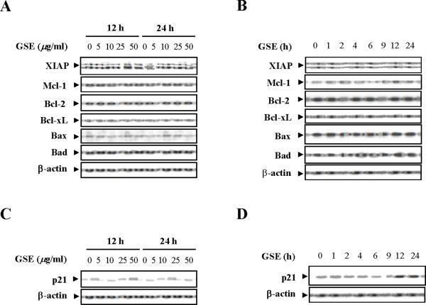
Figure 2.
GSE induces the expression of Cip1/p21 but does not affect the expression of Bcl-2 family members. (A) Jurkat cells were treated without or with various concentrations of GSE as indicated for 12 h and 24 h. (B) The cells were treated without or with 50 μg/ml GSE for 1, 2, 4, 6, 9, 12, and 24 h. For A and B, total cellular extracts were prepared and subjected to Western blot analysis using antibodies against Bcl-2 family members including XIAP, Mcl-1, Bcl-2, Bcl-xL, Bax, and Bad. (C) The cells were treated without or with the indicated concentrations of GSE for 12 h, and 24 h. (D) The cells were treated without or with 50 μg/ml GSE for 1, 2, 4, 6, 9, 12, and 24 h. For C and D, total cellular extracts were prepared and subjected to Western blot analysis using antibodies against Cip1/p21 (p21). For Western blot analysis, each lane was loaded with 30 μg of protein. Blots were subsequently stripped and reprobed with antibody against β-actin to ensure equivalent loading and transfer. Two additional studies yielded equivalent results.

