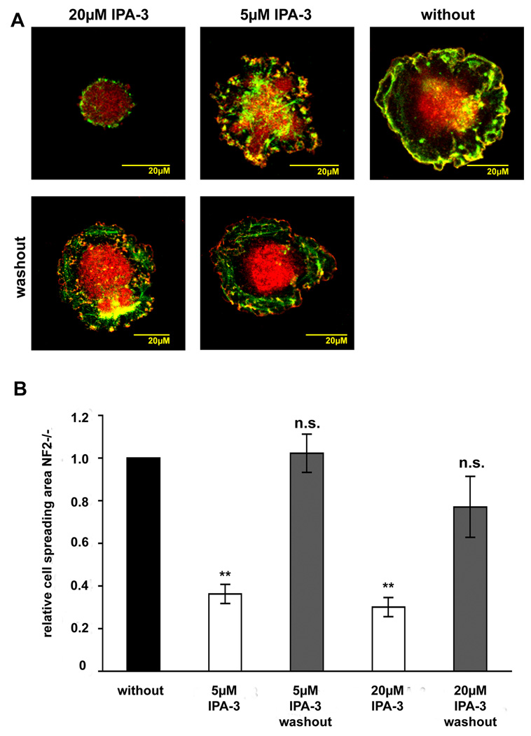
Figure 4.
Washout of IPA-3 in cell spreading experiments in human primary schwannoma cells (NF2−/−)
Schwannoma cells that were allowed to spread for 30 minutes in the absence or presence of the PAK inhibitor IPA-3 or the control substance PIR-3.5 on poly-L-lysine/laminin coated dishes were stained for the Arp2/3 complex (red) a marker for lamellipodia and ruffles and F-actin (green) to visualise cell morphology. IPA-3 was washed out after 30 minutes by changing inhibitor-containing media to inhibitor-free media. Cells were allowed to spread for another 30 minutes. Washout restored normal schwannoma cell morphology (A). Washout of 5 µM IPA-3 leads to the same cell spreading area compared to untreated cells and washout of 20 µM IPA-3 to an only slightly reduced cell spreading area that was clearly higher than in cells were inhibitor was left on (B).

