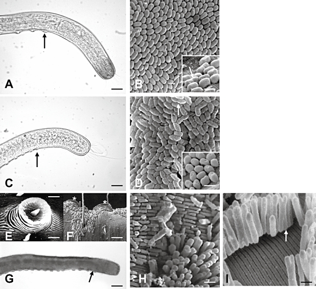
Fig. 1.

Photomicrographs of the anterior regions of fixed Robbea sp.1 (A), Robbea sp.2 (C) and Robbea sp.3 (G) and scanning electron microscopy (SEM) photographs of their respective symbionts (B, D, H and I). Black arrows point to the beginning of the suckers' row on each worm in (A), (C) and (G), while white arrows point to dividing symbionts in (B), (D) and (I). (E) and (F) are SEM photographs of one individual bacteria-free sucker, and two symbiont-coated suckers (asterisks) of Robbea sp.3, respectively. Scale bar is: 25 µm in (A) and (C); 1.5 µm in (B) and (D); 3 µm in (E); 8 µm in (F); 40 µm in (G); 2 µm in (H); 0.6 µm in (I).
