
Fig. 4.
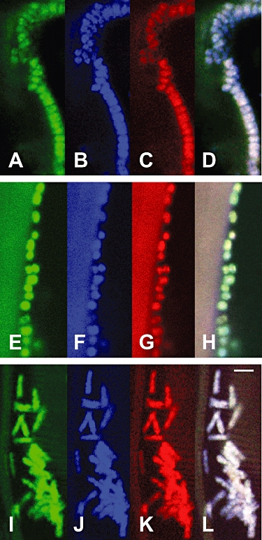
Fluorescence in situ hybridization (FISH) confocal microscope photographs of Robbea sp.1 (A–D), Robbea sp.2 (E–H) and Robbea sp.3 (I–L) symbionts attached to the worm surface. Each single symbiont is triple stained with a eubacteria-specific probe (green), a Gammaproteobacteria-specific probe (blue), and a symbiont-specific probe (red). (D), (H) and (L) are overlay pictures of (A)–(C), (E)–(G) and (I)–(K), respectively. Scale bar is 2 µm.