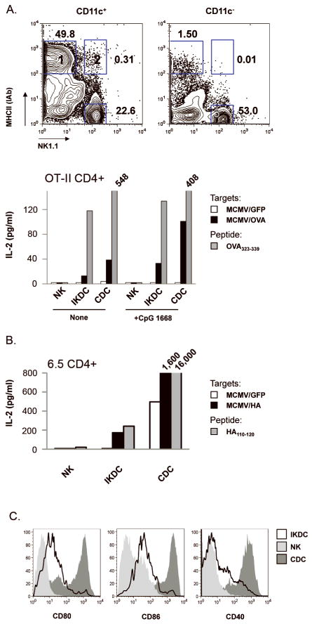
Figure 3. Upon recognition of MCMV-infected fibroblasts, IKDC differentiated into mature MHC-IIhi APC endowed with CD4+ T-lymphocyte stimulatory properties.
A, C57BL/6 CD11c+ and CD11c− splenocytes were incubated with MCMV/GFP- or MCMV/OVA-infected fibroblasts. CDC (CD11chiNK1.1−MHC-IIhi), IKDC (CD11c+NK1.1+MHC-II+) and NK (CD11c+NK1.1+MHC-II−) were FACS-sorted from gates 1, 2 and 3, respectively, and incubated with OVA-specific OT-II CD4+ T-cells. IL-2 was measured by ELISA in day 3 culture supernatants. ODN CpG 1668 was added or not to the culture to stimulate the APCs. APC were also incubated in presence of OVA323–339 to stimulate OT-II. B, Similar presentation assay was performed with HA antigenic model. The purity of sorted populations used in this assay is shown in Figure S3. C, Co-stimulatory molecules expression on CD11c+NK1.1+MHC-II+ IKDC (open) versus CD11c+NK1.1+MHC-II− NK (light grey) and CD11chiNK1.1−MHC-IIhi CDC (dark grey) in presence of infected fibroblasts.

