
Fig 6. Implant simulation and arch section module in InVivoDental.
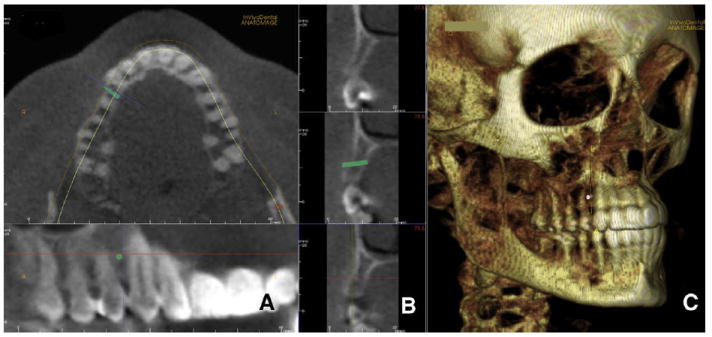
A, a microimplant is virtually placed between the roots of the maxillary right canine and first premolar; B, cortical bone thickness can be measured as well as total bone; C, InVivoDental also allows 3D visualization to assess anatomic relationships. The position on the implant can be modified with 6 degrees of freedom.