
Fig 7. Matching a 2D picture on the 3D soft-tissue volume.
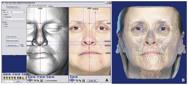
A, homologous landmarks are located in the volume and the 2D picture; B, Dolphin Imaging registers both images to create a multimodal image. Note the eye difference between the 2 modalities. There might be other, less-obvious areas of discrepancy between photographic and CBCT data.