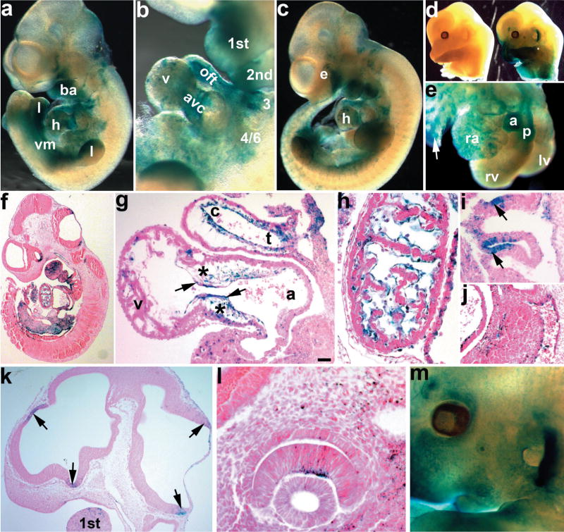
FIG. 1.
Analysis of X-gal stained E10.5 to E13.5 embryos from Smad7Cre; R26r indicator mice. Smad7Cre transgenic males were bred to R26r females. Wholemount X-Gal stained embryos were either photographed whole (a–d, m), partially dissected to expose heart (e), or serially sectioned and counterstained with eosin (f–l) to visualize Cre expressing cells. (a) Wholemount lacZ stained E10.5 Smad7Cre;R26r embryo. Note lacZ expression within the upper jaw, branchial arches (ba), ventral mesenchyme (vm), ventral region of the limb buds (l), and heart (h). (b) Higher magnification view of the first, second, third, and 4/6th branchial arches and heart in a, reveals lacZ-expressing cells in the outflow tract (oft) and atrioventricular cushions (avc), and punctate staining in the ventricles (v). (c) E11.5 Smad7Cre;R26r embryo shows more extensive expression in the craniofacial region, the eye (e), arches, heart, and limb buds. (d) E13.5 lacZ stained R26r only (left) and Smad7Cre;R26r (right) littermate embryos. No expression is seen in R26r only embryos when Cre is absent, verifying tissue-restricted reporter expression. (e) Isolated E13.5 heart from Smad7Cre;R26r embryo. Note robust lacZ expression in both aorta (a) and pulmonary (p) vessels, as well as punctate lacZ in right atria (ra) and left and right ventricles (lv,rv). Also note lacZ expression in bronchi of nascent lungs (arrow). (f) Eosin counterstained paraffin section of the embryo in a. (g) Sagittal section of E10.5 Smad7Cre;R26r embryo heart showing robust lacZ expression within both the OFT and AV mesenchymal cushions (*) and overlying endocardial cell (arrows) lineages. Note lacZ is present within both the conus (c) and truncus (t) of the OFT. (h) E10.5 sagittal section through the myocardium of the heart, note lacZ is con- fined to the endocardial cells and absent from the cardiomyocytes. (i) E10.5 sagittal section through aortic arch arteries, showing robust lacZ expression in the ectodermal pouches. (j) E10.5 section through the aorta showing lacZ staining is restricted to the endothelium (arrow). (k) Sagittal section of the E11.5 Smad7Cre;R26r embryo in c, showing lacZ in the neuroepithelium of the forebrain and hindbrain (arrows) and within the first branchial arch. (l,m) High power section of lacZ in E11.5 inner neural layer of the optic cup (l), and low power wholemount view of lacZ expression in E13.5 eye and ear (m). Scale bar in g = 20 μm. Abbreviation: v = ventricle.

