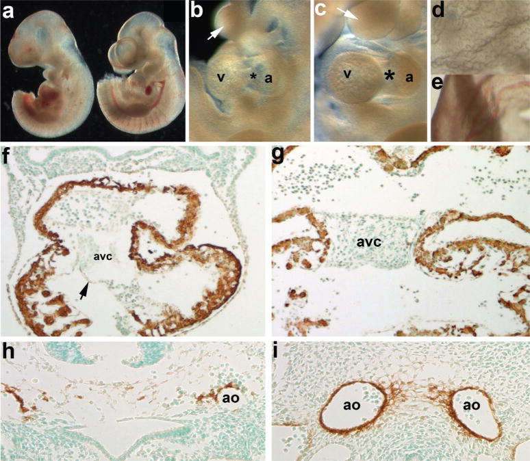
FIG. 2.
Evaluation of resultant cardiovascular phenotypes following Cre/loxP-mediated genetic cell ablation in Smad7Cre;R26DTA embryos. (a) E10.5 Smad7Cre;R26DTA (left) and R26DTA only (right) embryo littermates. (b,c) Higher power images of ablated mutant (b) and normal control (c) cardiovascular and branchial arch regions of embryos shown in a. Note that the AV cushion region (*) is largely absent in mutant (b) hearts and that the arches are smaller in size (arrows). (d,e) High power images of the mutant (d) and normal control (e) yolk sacs, note the lack of vasculature and blood in mutant. (f) Counterstaining with αSMA (that detects both smooth and cardiac muscle lineages at this stage of in utero development), reveals that the myocardium is largely unaffected via Smad7Cre-mediated cell ablation but that the overall size of the heart is decreased, when compared to unaffected littermate photographed at same magnification (g). Also note that while the endocardial cushions are hypoplastic, the mutant endothelium remains intact (arrow, g). (h,i) α-SMA staining also reveals that whilst the control littermate has two distinct α-SMA-positive paired dorsal aorta (i), and the ablated mutant vessels have hemorrhaged and that there is fragmented α-SMA-stained smooth muscle cells within the mutant (h). Sections f–i were lightly counterstained with methyl green. Abbreviation: a = atria.

