Figure 1.

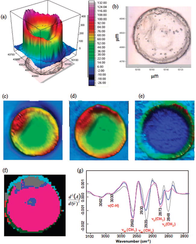
High-resolution synchrotron FT-IR map of a GV cell recorded using a 4 μm × 4 μm aperture: (a) Total absorbance map constructed by integrating the area between 1800 and 1000 cm−1. (b) Photomicrograph of a GV cell showing the clear nucleus and scale bar. (c) Chemical map of the integrated area under the amide I band. (d) Chemical map of the integrated area of the CH2 and CH3 stretching region (3000−2800 cm−1). (e) Chemical map of the integrated area of the ester carbonyl band (1720−1750 cm−1). (f) False color five cluster map generated by performing UHCA on the CH stretching region (3100−2800 cm−1). (g) Mean extracted spectra from the blue, gray, and pink clusters.
