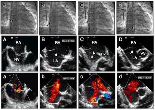
Figure 1.

Fluoroscopic and intracardiac echocardiographic images during transcatheter closure of a large secundum atrial septal defect in a 12 year old male child. A, A, a position of the ICE catheter (arrow) during home view. B, B, b images in septal view without and with color Doppler demonstrating the presence of a large defect (arrow) measuring 22 mm. C, C, c, images in caval (long axis) view demonstrating the defect (arrow) and the rims. D, D, d, images in short axis view demonstrating the defect (arrow) and absence of anterior rim. RA: right atrium; RV: right ventricle; LA: left atrium; SVC: superior vena cava.
