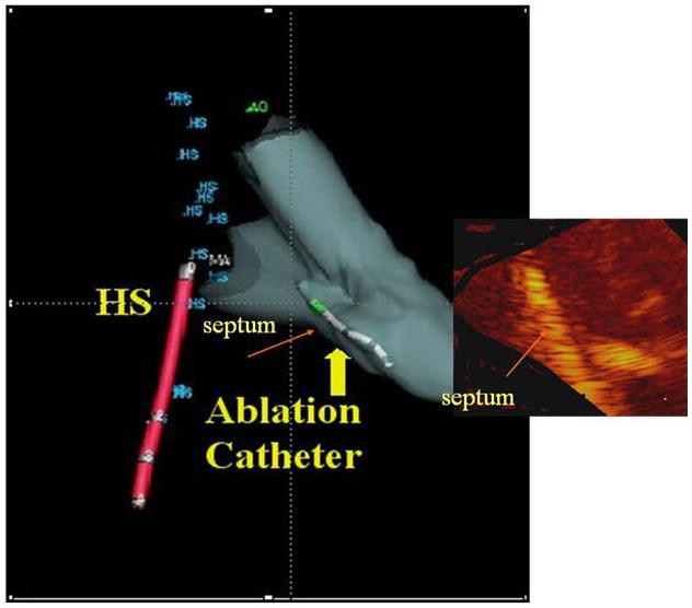
Figure 10.

Fusion image showing the ultrasound image of ablation overlaid onto the NavX (St. Jude Medical) shell with a retrograde ablation catheter ablating the left side of the septum, which has brightened. This also requires reading the rotational position of the side-looking Hockeystick catheter into the electrofield NavX (St. Jude Medical) milieu.
