Fig. 4.

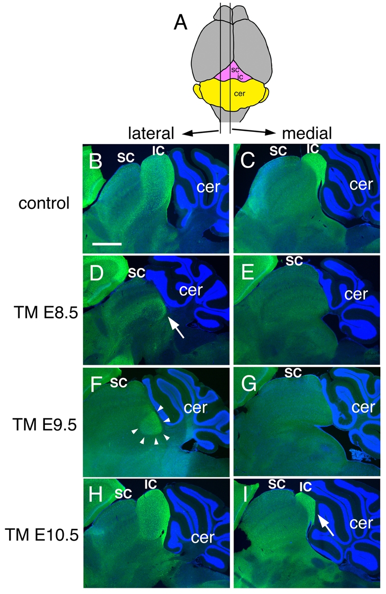
Neurogranin staining of adult brain sections shows that the inferior colliculus requires sustained Fgf8 signaling. (A) Schematic of dorsal view of an adult brain to show the level of the lateral and medial sections shown below. (B-I) Neurogranin staining of the inferior colliculus of adult Fgf8 temporal CKO embryos treated with TM at the time-points indicated. The control is an Fgf8flox/+; En2CreER/+ embryo treated with TM at E8.5. The arrow in D indicates the faint and abnormally positioned neurogranin staining in the Fgf8-E8.5 CKO. Arrowheads in F indicate the abnormal neurogranin staining in the lateral IC of a Fgf8-E9.5 CKO mutant. Arrow in I indicates the smaller medial IC in Fgf8-E10.5 CKO mice. Abbreviations are as in Fig. 1. Scale bar: 500 μm.
