Fig. 5.
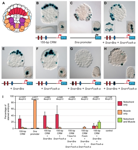
Requirement of both Ci-Bra and Ci-FoxA-a for ectopic activation of the 155-bp notochord CRM. (A) Simplified lineage map of the vegetal half of the Ciona embryo at the 110-cell stage. Notochord precursors are labeled in red, muscle precursors in orange, trunk mesenchyme in purple and neural precursors in light blue. Precursors of two different lineages are labeled with two colors. (B-H) Embryos electroporated at the one-cell stage with the constructs indicated underneath each panel. Binding sites that have been mutated are covered by an `X'. Embryos were cleavage arrested with cytochalasin B at the 110-cell stage and stained for β-galactosidase (blue). In all panels, the insets at the bottom right show embryos treated with cytochalasin B at the early gastrula stage. (B) Activity of the 155-bp Ci-tune CRM. Bottom left inset shows an untreated control embryo from the same clutch, grown in parallel with the cleavage-arrested embryos shown in all panels. (C) Activity of the Ci-Sna promoter, which was used to drive misexpression of Ci-Bra and Ci-FoxA-a. (D) Triple electroporation of the 155-bp CRM with misexpression constructs for Ci-Bra (Sna>Bra) and Ci-FoxA-a (Sna>FoxA-a) showing ectopic muscle staining only on one side, due to mosaic incorporation. Bottom left inset depicts another embryo from the same experiment, showing bilateral muscle staining. (E) Co-electroporation of the 155-bp CRM with the Sna>Bra misexpression construct. (F) Co-electroporation of the 155-bp CRM with the Sna>FoxA-a misexpression construct. (I) Graph showing the percentage of cleavage-arrested embryos with staining in notochord (red bars), muscle (orange bars), or in both tissues (green bars). The transgenes employed are indicated underneath the x-axis.

