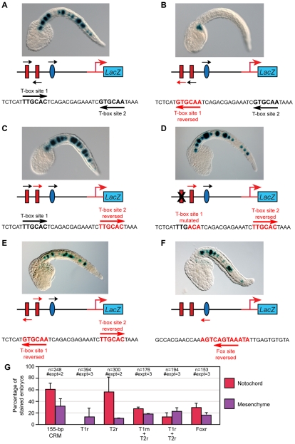
Fig. 6.
Effects of changes in the orientation of the Ci-Bra and Ci-FoxA-a binding sites on notochord activity of the Ci-tune CRM. Late-tailbud embryos electroporated at the one-cell stage with either the wild-type 155-bp Ci-tune CRM or with one of its mutant versions, schematized underneath each microphotograph. Black arrows indicate the native orientation of the sites, red arrows indicate the new orientation created by mutagenesis. Embryos are stained for β-galactosidase (blue). (A) In the wild-type Ci-tune CRM, the cores of T-box site 1 and T-box site 2 are identical, but are oriented in opposite directions. (B) Reversal of T-box site 1. (C) Reversal of T-box site 2. (D) Reversal of T-box site 2, combined with a mutation of T-box site 1. (E) Reversal of both T-box sites. (F) Reversal of the Ci-FoxA-a site. (G) Quantification of notochord and mesenchyme staining following electroporation with the wild-type 155-bp CRM and with its derivatives in which the Ci-Bra and Ci-FoxA-a sites are reversed and/or mutated. Embryos were scored as in Fig. 2. T1r, T-box site 1 reversed; T1m, T-box site 1 mutated; T2r, T-box 2 site reversed; Foxr, Fox site reversed.

