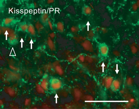
Figure 12.
Expression of PR in Kiss1 neurons. Photomicrograph of cells costaining for kisspeptin (green) and PR (red) in the ARC of the ewe brain. Arrows indicate cells containing both kisspeptin and PR. The open arrowhead indicates a kisspeptin-positive PR-negative cell. Scale bar, 50 μm. [Modified with permission from Smith et al., 2007 (107) © The Endocrine Society].

