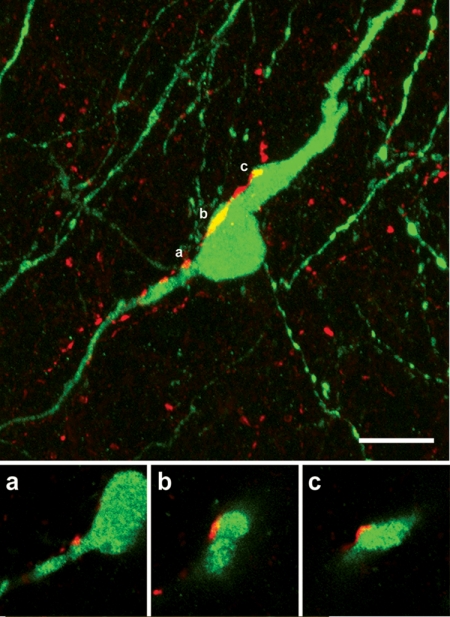
Figure 6.
Kisspeptin projections to GnRH neurons in adult female mice. Confocal stack of 75 images showing a single GnRH neuron (green) with kisspeptin (red) fibers surrounding and apposed to it. Single 370-nm-thick optical sections through the three regions indicated by a, b, and c of the GnRH neuron are given below to demonstrate the close apposition between kisspeptin fibers and GnRH neuron elements. Scale bar, 10 μm. [Modified with permission from Clarkson and Herbison, 2006 (73) © The Endocrine Society].

