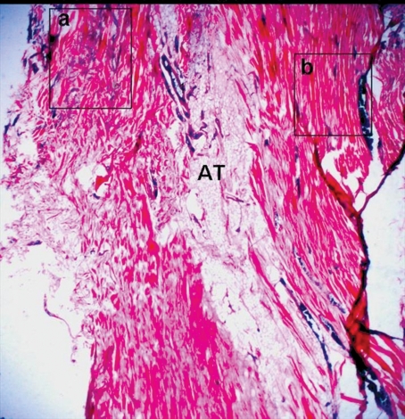
Figure 3.
Microphotograph of a specimen containing a unit of SML-DML. a. The DML is running at the left section. Elastic fibers are supernumerary in the “a” square b. The SML consisted of collagen fibers is indicated by the “b” square. Two ligaments are separated with an areolar adipose tissue (AT) (X2.5).

