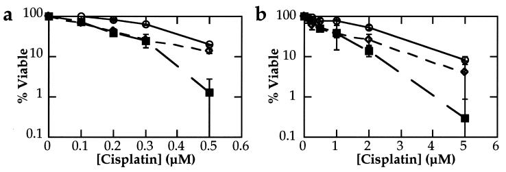
Figure 5.
Colony formation assays. The p53-wild-type F9 (○) and Nulli-SCC1 (◊) and p53-mutant EB16 (▪) teratocarcinoma cell lines were treated either continuously (a) or for 2 h (b) with cisplatin. After incubation for 5–7 days, visible colonies were stained with methylene blue and counted. For each cell line, the numbers were normalized to the untreated data point. Bars ± 1 esd.

