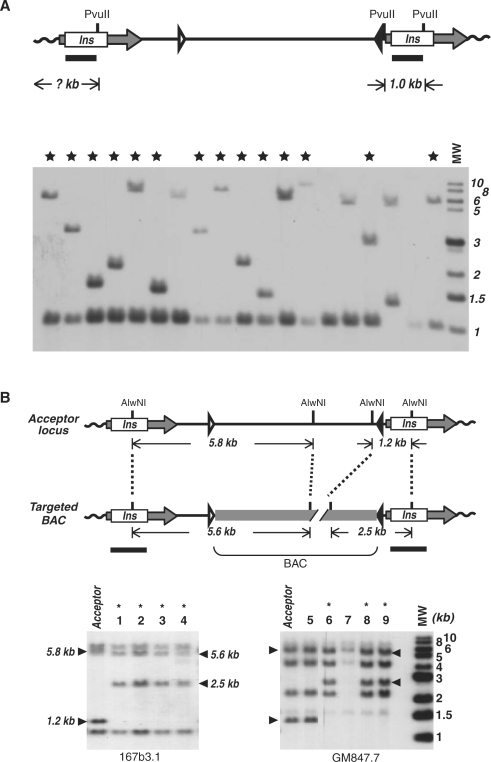
Figure 3.
Southern analyses of acceptor loci and targeted BAC integrations. (A) Analysis of proviral acceptor loci. Upper panel is a diagram of a proviral acceptor locus following retroviral transduction. Horizontal arrows indicate defective LTR elements in which the U3 regions are replaced by the insulator cHS4 (Ins). Lox511 and loxP sites are shown as open and closed triangles, respectively. Lower panel shows a representative Southern analysis of acceptor loci. Genomic DNAs were digested with Pvu II and hybridized to a 32P-labeled Pvu II-Hind III fragment of cHS4. MW, molecular weight markers in kilo base pairs. Stars mark the acceptor clones with a single-copy provirus. (B) Analysis of integrated BAC constructs. Upper panel illustrates an acceptor locus before and after targeted BAC integration. The integrated BAC is shown as a gray thick line. Sizes of internal genomic bands recognized by the insulator probe are also indicated. Lower panels show representative Southern analyses of BAC constructs integrated into acceptor loci in 167b3.1 (left) and GM847.7 (right) cells. Genomic DNAs were digested with AlwN I and hybridized to a full-length insulator probe. As indicated by arrowheads, the 5.8-kb and 1.2-kb internal bands changed to 5.6 kb and 2.5 kb, respectively, upon integration of the BAC, whereas the two external bands specific to each acceptor locus were not affected. The positions of the Southern probe are shown as horizontal bars below the diagram. Asterisk indicates clones with targeted integration of the BAC reporter.

