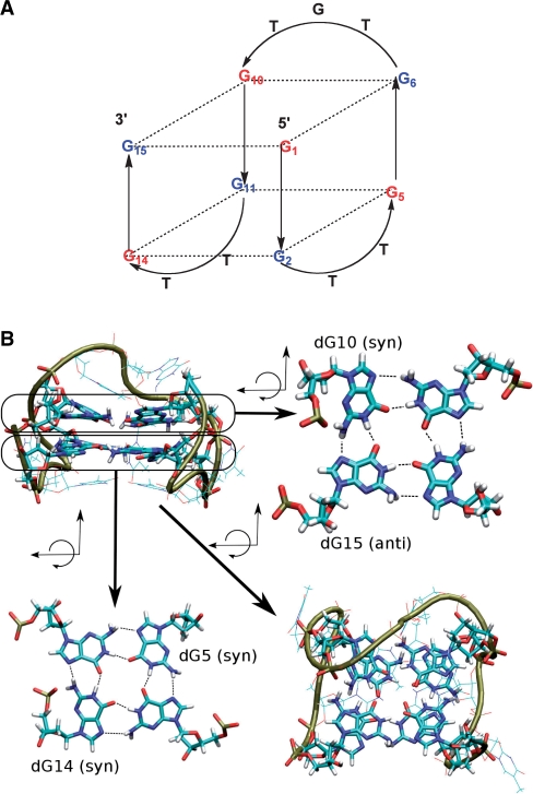
Figure 1.
(A) Schematic representation of the antiparallel folding of the thrombin-binding aptamer (TBA); blue represents guanines that are anti and red guanines that are syn. Positions of 5, 10, 14 and 15 were modified in this study. (B) Molecular representation: the upper left quadrant shows a lateral view of the TBA and the lower right quadrant a view from the top of the molecule [dG5(syn) and dG14(anti) in the lower quartet as well as dG10(syn) and dG15(syn) in the upper quartet are indicated].

