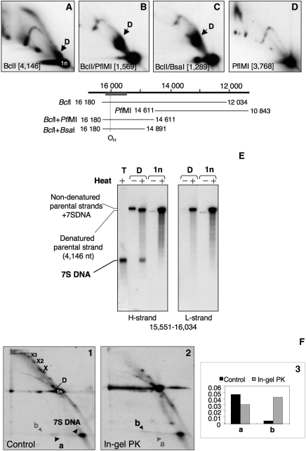
Figure 2.
Both molecular species depleted by POLGβ siRNA contain 7S DNA. Restriction fragments of mouse mtDNA containing the D-loop region were visualized by hybridization with a radiolabelled probe after separation by 2D-AGE (A–C); all had a product equivalent to species D of human mtDNA (Figure 1C), and this was absent from fragments lacking the D-loop region (D). The co-ordinates of the restriction fragments analysed in (A–D) are illustrated schematically immediately below the 2D gel images. (E) Extraction of species D after 2D-AGE of BclI digested mouse liver mtDNA (A) and subsequent heat denaturation released 7S DNA, which was resolved and detected by 1D-AGE and Southern hybridization. 7S DNA was present in undigested mouse mtDNA samples (T), but was not associated with the unit length BclI fragment (1n) spanning nt 12 034–16 180. (F) A substantial amount of 7S DNA is released following restriction enzyme digestion of HOS cell mtDNA (gel image 1). Species D is presumed to be present (based on previous analysis (26), but was not clearly separated from the unit length fragment (1n) under the conditions used here. Species ‘a’ has the same mobility as 7S DNA in the second dimension but not the first; therefore it is inferred to be 7S DNA that is released between the first and second dimension electrophoresis steps. Another species with the same mass as 7S DNA (b) is released from molecules resolving close to the apex of the X arc when the same material is treated with proteinase K between the first and second dimension gel electrophoresis steps (gel image 2). (Image 3) phosphorimager quantification of species a and b. Note that the species at the apex of the X-arc may contain 7S DNA, but if so, its stability is not dependent on the presence of protein.

