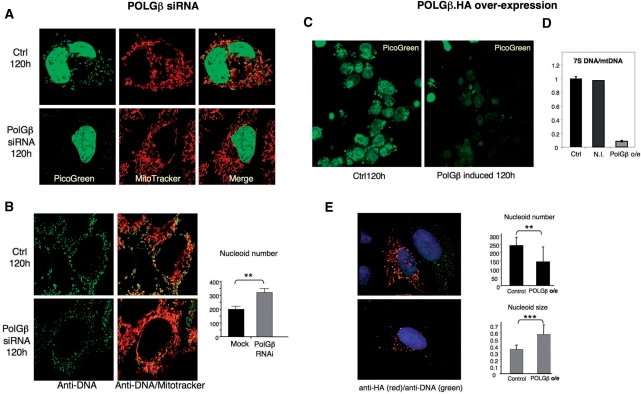
Figure 4.
Modulating the expression of POLGβ affects mitochondrial nucleoid structure, number and size. HOS cells displayed a substantial decrease in PicoGreen staining of mitochondrial nucleoids after POLGβ siRNA (A and Supplementary Figure 1). Cells were twice mock-transfected (control), or transfected with dsRNA-840-targeting POLGβ (POLGβ RNAi). Transfections were at t = 0 and t = 72 h; live cell imaging was performed at t = 120 h. DNA was stained with Picogreen and mitochondria with Mitotracker™ (Invitrogen); both reagents depend on a membrane potential for mitochondrial uptake. (B) Mock and POLGβ siRNA treated HOS cells were immunolabelled using anti-DNA antibody, visual inspection of cells suggested that nucleoids were more numerous after POLGβ siRNA and this was confirmed by analysis of 29 cells of each type from three independent experiments with Axiovision software (Zeiss): P = 0.004 based on the unpaired two-tailed student's t-test (**P < 0.01). Control and dsRNA-840 treated cells with the most intense signal were selected for nucleoid counting from several different fields of cells, as such cells were invariably well spread and in good condition. Induction of POLGβ transgene in HEK cells for 120 h led to a loss of Picogreen signal (C) and a marked decrease in 7S DNA level after 72 h treatment with 10 ng/ml doxycyclin, compared to the same cells not induced (N.I.) with the drug and control (Ctrl) HEK cells lacking the POLGβ transgene (D). (E) Over-expression of HA-tagged POLGβ in human U2OS osteosarcoma cells resulted in the formation of enlarged nucleoids (stained with anti-DNA antibodies) (**P < 0.01), which were fewer in number than in untransfected cells (***P < 0.001) (see ‘Results’ section and Supplementary Figure 3). U2OS cells were preferred to HEK cells for confocal analysis because they spread well and therefore yielded better quality images.

